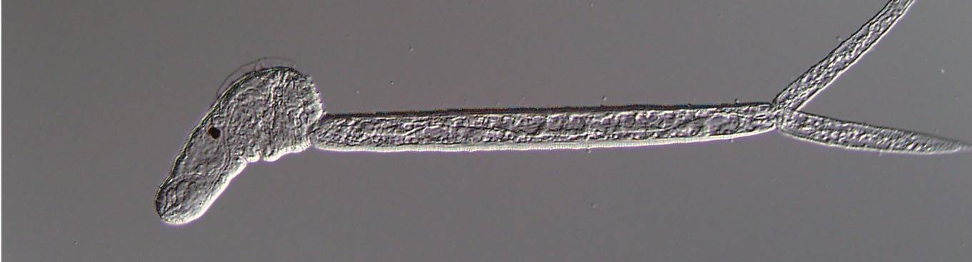
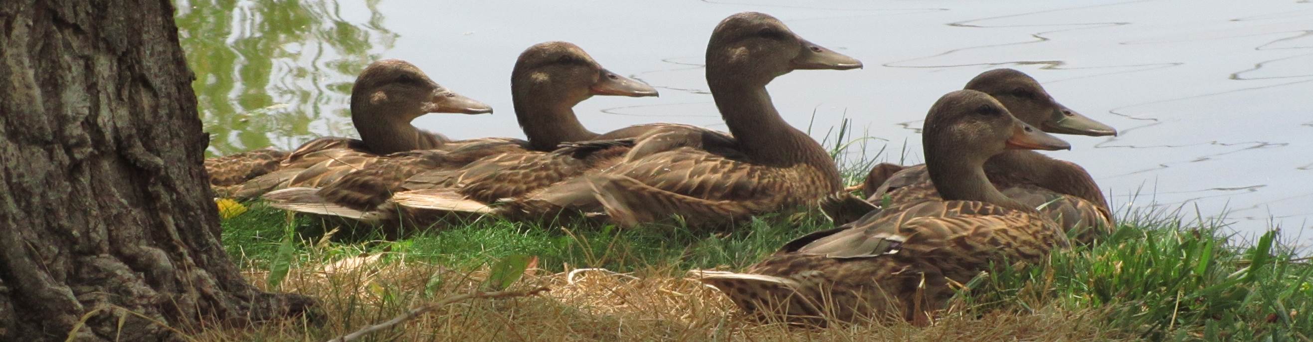
Life Cycle: The life cycles of the Allassostomoides genus are not well known. However the life cycle of A. parvum was experimentally determined to utilize snail hosts where rediae produce cercariae (Beaver 1929). Cercariae encyst on the shells of snails, skin of frogs, or vegetation (Olsen 1974). Frogs and turtles become infected with the adult (Olsen 1974).
Description:
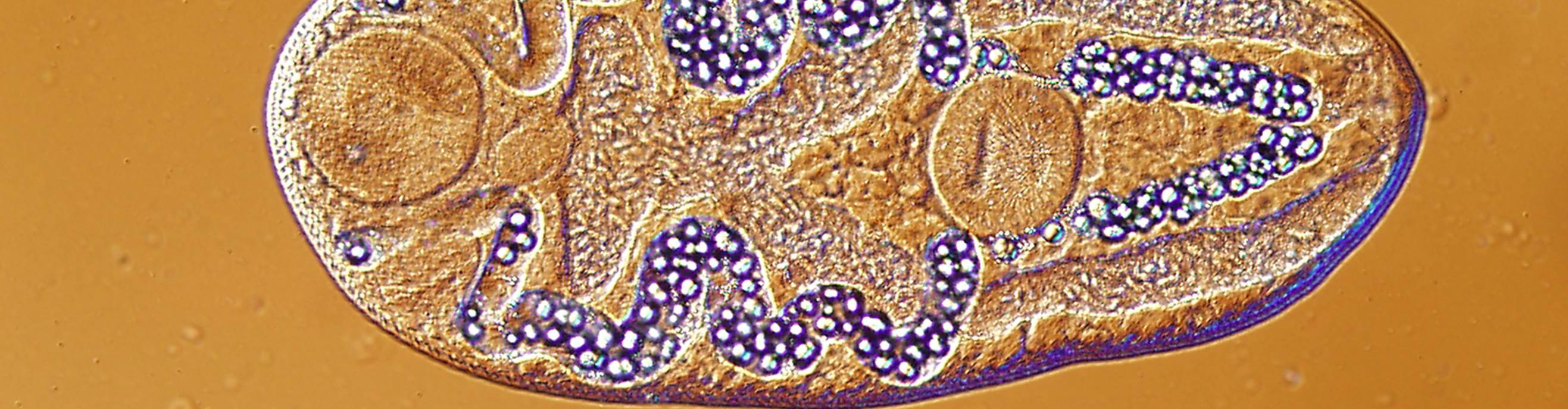
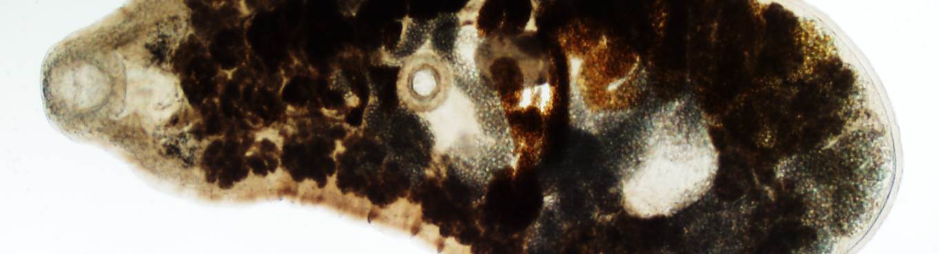
The body is capable of considerable elongation and in a fully extended specimen the two lateral prominences that ordinarily are present on either side of the acetabulum almost disappear. In the different specimens there is a large amount of variation in size but otherwise such morphological agreement that I am convinced they belong to the same species. They vary from 3 to 5.5 mm in length and from 0.8 to 1.6 mm in width. The acetabulum measures from 0.67 to 1.2 mm in length and from 0.64 to 1 mm in width. The oral sucker is oval. It varies from 0.32 to 0.64 mm in length and from 0.27 to 0.46 mm in width. The details of its structure were described in the original account. The oral evaginations are visible in whole mounts; they arise from the dorsal posterior lateral margins of the sucker and measure from 0.06 to 0.08 mm in length. The course of the esophagus depends on the condition of the anterior end of the body and in a protracted state the esophagus is a straight tube. In length it varies from 0.3 to 0.6 mm and in width from 0.035 to 0.07 mm throughout its length it is enclosed by deeply staining gland cells. This organ may be spherical or oval, but in well extended specimens it is longer than broad. It varies from 0.16 to 0.3 mm in length and from 0.14 to 0.21 mm in width. At the posterior end of the esophageal bulb on either side there is a short tube which extends obliquely laterad and posteriad. These short tubes are lined with cuticula continuous with that of the esophagus and form a bridge across the median plane of the body separating the intestinal diverticula. Their diameter is about one-fifth that of the ceca and their length is about equal to their diameter. The ceca arise from their caudal ends and expand immediately into large sack-like tubes almost one-fourth of the body width in diameter, which extend posteriad and terminate just anterior to the acetabulum. The testes are contiguous, one before the other, in the anterior half of the worm. They are much farther forward in this species than in A. magnum. They are situated almost in the median plane, although in some specimens there seems to be a tendency for one to lie slightly at the right and the other slightly at the left of the midline. These organs are spherical to oval in shape and measure from 0.28 to 0.32 mm in length and from 0.24 to 0.32 mm in width. The primary ducts, seminal vesicle, and cirrus sac have been described and no addition to that account is necessary. In these larger worms the reproductive organs have attained maturity and increased functional activity. Consequently the structures are larger and the seminal vesicle is conspicuous in whole mounts. The ovary is spherical to oval, longer in either the antero-posterior or lateral axis, and measures from 0.16 to 0.2 mm in diameter. It is situated in or near the median plane, slightly posterior to the middle of the body. The oviduct arises at the posterior margin and passes posteriad where it enters the ootype. The female genital complex in the present worms agrees in essential features with the description of the immature specimens. Mehlis' gland is large, and after the entrance of the vitelline duct the uterus passes forward. It may lie on either side of the ovary and often the latter organ is pushed slightly left or right of the median plane by the uterus which fills the opposite side of the body. Throughout its course the uterus is filled with large oval eggs which average 0.145 mm in length and 0.1 mm in width. The uterus in adult specimens is much coiled and as many as fifty eggs have been counted in a single worm. The vitelline follicles extend from the level of the anterior testis almost to the ends of the digestive ceca. Anteriorly they are lateral in position, behind the testes they enter the intercecal area but do not extend to the median plane, and behind the ovary they become confluent. The follicles are very large, spherical, oval, or triangular in outline, and in diameter they measure from 0.1 to 0.2 mm. Ducts from the two sides join behind the ovary and discharge into the ootype.
Sources: Beaver, P.C. 1929. Studies on the Development of Allassostomoides parvum Stunkard. Journal of Parasitology, Vol. 16, p. 13-23.
Olsen, O.W. 1974. Animal parasites, their life cycles and ecology. University Park Press, London, Baltimore and Tokyo, 3rd Revd Edn., p. 278-279.